(Note this is part 2 of a 2 part series of posts on Visual Processing)

An illustration of various types of stimuli and their evoked responses by different visual cortices can be seen in a study on visual processing of shapes by Joakim Vinberg. Vinberg created stereoscopic and moving images of an object (a star), a hole in a surface, edges, surfaces and random noise. He then measured the response of different visual cortices, V1, V2, V3, V3a V4 and MT, to each of the stimuli.[iii]
The study revealed that the MT area was least responsive area tested for the stereo presentation of a hole, while the V1 area was most responsive. Similarly The V3 area responds more strongly to random moving stimuli than the V3a area.

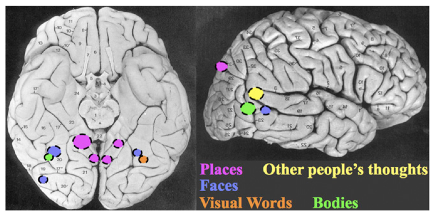
Some striking example of targeting specialized functional cortical regions is demonstrated by the work of Nancy Kanwisher.[iv] Using fMRI Kanwisher has identified cortical regions that consistently respond to specific categories of visually perceived objects. The regions identified respond twice as strong to their preferred stimuli as they do to any other type of object.
For example a small area of the bottom surface of the cerebral cortex, which is activated when a face is perceived.[v] This area responds to face-like arrangements of eyes, noses and mouths and also things like cartoon images of faces. Its perception is size, location and viewpoint of the face-like stimuli.
Another identified area adjacent to the collateral sulcus in parahippocampal cortex is activated when presented with any scene type of layout. The scene may be indoor or outdoor, but seems to require spatial information being presented.
An area adjacent to the visual motion area MT is activated by images of other people’s bodies or body parts. It even responds to images of the bodies of animals and stick figures.
And finally a very small region was shown to respond to stimuli that were written words and strings of consonant letters, but did not activate for numerical digits, line drawings or letters from unfamiliar alphabet systems.
This ability to create a more finely tuned response, along with wide bandwidth are great advantages to VBCI. Together they may be used to create a robust, fast and noninvasive means of communicating information from computers into the human mind.
[i] “Rabbit–duck illusion,” Wikipedia, en.wikipedia.org/wiki/Rabbit-duck_illusion, (April, 26, 2017).
[ii] M. S. Gazzaniga, R. B. Ivry, and G. R. Mangun, Cognitive Neuroscience: The Biology of the Mind, 3rd edition (W. W. Norton, 2015) 408.
[iii] Joakim Vinberg1 and Kalanit Grill-Spector, Representation of Shapes, Edges, and Surfaces Across Multiple Cues in the Human Visual Cortex (J Neurophysiol 99: January, 2008) 1380–1393.
[iv] Kanwisher, N. (2010). Functional specificity in the human brain: A window into the functional architecture of the mind. Proceedings of the National Academy of Sciences, 107(25), 11163-11170. doi:10.1073/pnas.1005062107
[v] Kanwisher , “Functional specificity,” 11164.